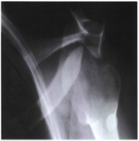
GARTH METHOD
Pathology Demonstrated:
An optimal trauma projection for possible scapulohumeral dislocations (especially posterior dislocation), glenoid fractures, Hill-Sachs lesions, and soft tissue calcifications.
Technical Factors:
- IR size - 18 x 24 cm (8 x 10 inches), lengthwise
- Moving or stationary grid
- Digital IR - very close collimation required
- 75 +- 5 kV range
- mAs 12
Shielding:
 |
Erect apical oblique axial projection - 45 degrees posterior oblique
CR 45 degree caudad |
Shield pelvic area.
Patient Position:
Perform radiograph with the patient in an erect or supine position. (The erect position is usually less painful, if patient's condition allows.) Rotate body 45 degree toward affected side ( posterior surface of affected shoulder against IR.)
Part Position:
Center scapulohumeral joint to CR and mid-IR.
Adjust IR so that the 45 degree angled CR will project the scapulohumeral joint to the center of the IR.
Flex elbow and place arm across chest, or with trauma, place arm at side as is.
Central Ray:
CR 45 degree caudad, centered to the scapulohumeral joint.
Minimum SID of 40 inches (100 cm)
Collimation
Collimate closely to area of interest.
Respiration:
Suspend respiration during exposure.
Radiographic Criteria:
 |
AP axial oblique: Garth method demonstrates an antrior dislocation of the
proximal humerus. The humeral head is shown below the coracoid process,
a common appearance with anterior dislocation |
Structure Shown:
The humeral head, glenoid cavity, and neck and head of the scapula are well demonstrated free of superimposition.
Position
The coracoid process is projected over part of the humeral head, which appears elongated.
The acromion and AC joint are projected superior to the humeral head.
Collimation and CR:
Collimation should be visible on four sides to area of affected shoulder.
CR and center of collimation field should be at the scapulohumeral joint.
Exposure Criteria:
Optimal density and contrast with no motion will demonstrate clear, sharp bony trabecular markings and soft tissue detail for possible calcifications.


