Anal Fistula MRI Planning
Perianal Fistula is an abnormal attachment between the epithilialised surface of the anal canal and the skin. There are two main causes of this conditions. The primary condition is an obstruction of anal gland which leads to stasis and infection with abscess and fistula formation and this is the most common causes. The secondary cause are enumerated below:
- Iatrogenic - hemorrhoideal surgery
- Inflammatory bowel diseases - Crohn's disease more common than colitis ulcerosa
- Infection - viral, fungal or TB
- Malignancy
Patient Position
After the safety check have been performed, position the patient supine on the scanning table with their heads on the pillow. Give the patient an emergency buzzer and provide head phone to protect the patients ears. Place the body coil across the patient’s pelvic area from the iliac crest downwards.
Secure the coil, move the patient into the scanner and center the lazer beam localizer into the middle of the coil.
Move the patient fully in into the coil making sure that they are calm and comfortable before leaving the room. Once you are in the control room select the correct patient details from the browser or you can type the details manually.
It’s very important to get the details right, including the correct patient weight so that the SAR or Specific Absorption Rate can be calculated accurately. Register the patient as lying head first and supine, select the correct protocol according to your hospital and radiologist guidelines.
Localizer Sequence
T2 Sagittal High Resolution of the Pelvis
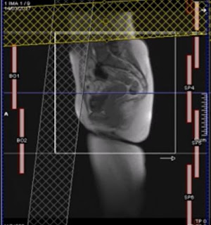
To start your scanning, run a localizer sequence in 3 planes the sagittal, coronal and the axial planes.
Sagittal Localizer
After scanning the localizer, run the localizer plan on your first sequence which is usually a sagittal T2 high resolution of the pelvis. When planning on the sagittal localizer it is very important to make sure that the entire soft tissue of the buttock is included in the field of view (FOV). Saturation band above and in front of the field of view will reduce artifacts from ball motion and breathing.
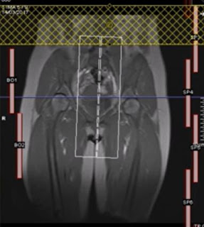
Coronal Localizer
On the coronal localizer align the central slice to the midline of the body through the symphysis pubis.
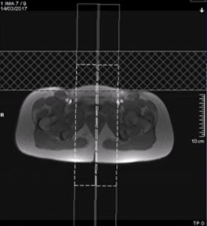
Axial Localizer
Routine Sequence
Fat saturated T2 sagittal Sequence
You can copy your planning from the previous sequence.
Check if the covered of the planning is sufficient then select apply.
High Resolution T2 axial Fat Sat
Once your planning of your first sagittal is complete you can plan your high resolution T2 axial fat sat, slices should run perpendicular to the anal canal, and its very important to cover from the mid rectum right down to the skin surface of the buttock as fistuli contract through the tissue to open through the skin, check your planning if straight and center the coronal plane likewise to the axial plane and apply.
High Resolution T2 coronal Fat Sat Sequence
Now plan a high resolution T2 coronal fat sat sequence, this time your slices will run parallel to the anal canal. The planning box should cover the skin suface of the buttock, straighten the other plains and make sure the body coil are turn on then apply.
 |
| HR T2 Coronal Planning |
Reviewing Images
T2 Sagittal Images
In T2 sagittal images where fluids and fats appear bright this particular study, clearly visualize the perianal fistula beginning to the anal canal and tracking to the surface of the skin.
T2 fat saturated images
Where fluids appear bright and fat appears dark in T2 fat saturated sequences fistula track are better visualize due to the nullified signal of the fat.
 |
| T2 Fat Saturated |
 |
| T2 Fat Sat - Fistula track (arrow) |
T2 Fat saturated Axial
This view of the anal canal is the one most important sequences in fistula imaging. In this axial image you track the fistula from anus sphincter down to the subcutaneous tissue.
 |
| T2 Fat Saturated Axial |
T2 fat saturated Coronal


