Radio Imaging Plane
Understanding the intricacies on radiography specially on CT scanning and MRI requires the familiarity with imaging planes. One of the example is the bread analogy which help explain the body planes. This is a brief review of the directional terms used in medicine may also make a discussion of the body planes easier to understand.All directional terms are based on the body being viewed in the anatomic position. This position is characterized by an individual standing erect, with the palms of the hands facing forward.
 |
| Anatomical Position |
This position is used internationally and guarantees uniformity in descriptions of direction.
Radiographic Positional Terms
The term anterior and ventral refers to movement forward or towards the face. Posterior and dorsal are equivalent terms used to describe movement toward the back surface of the body.
Inferior refers to movement toward the feet or down and is synonymous with caudal or toward the tail or, in humans, the feet. Superior defines movement toward the head and is used interchangeably with the term cranial or cephalic. Lateral refers to movement toward the side of the body. Inversely, medial refers to movement toward the midline of the body.
The term distal and proximal are most often used referring to extremities or limbs. Distal or away form, refers to movement toward the ends. The distal end of the forearm is the end to which the hand is attached. Proximal or close to, which is the opposite of distal, may be defines as situated near the point of attachment. For example, the proximal end of the arm is the end at which is attaches to the shoulder.
To help visualize the imaginary body planes, it is helpful to think of large sheets of glass cutting through the body in various ways. All sheets of glass that are parallel to the floor are called horizontal, or transverse planes. Those that stands perpendicular to the floor are called vertical, or longitudinal planes.
 |
| Imaging Planes |
When a sheet of glass will divides the body into anterior and posterior sections is the coronal plane. The sagittal plane divides the body into right and left sections. The sagittal plane that is located directly in the center, making left and right sections of equal size, is appropriately referred to as the median, or midsagittal plane. A parasagittal plane is located to either the left or the right of the midline. Axial planes are cross sectional planes that divides the body into upper and lower sections. Oblique planes are planes that are slanted and lie at an angle to one of the 3 standard planes
 |
| Oblique planes lie at an angle to one of the standard planes. |
Changing the image plane shows the same structures in a new perspective. The loaf of bread analogy can help to explain this change. Like for example if a coin is baked within the bread and lies standing on edge in the loaf, a sharp knife cutting through the bread lengthwise will show the coin as a flat, rectangular density. However, if the bread is restacked and cut in an axial plan, the coin appears circular.
 |
| The imaging plane will affect the way that an area of anatomy is represented on the cross-sectional slice. |
The image plan can be adjusted by positioning the patient, gantry or both to permit scanning in the desired plane or by reformatting the image data. Scanning in the desired data, although advances in CT technology have reduced the quality difference.
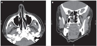
Changing the image plane in CT provides additional information in a fashion similar to the coin within the bread. Changing the image plane from axial to coronal is indicated for two distinct reasons. The primary reason is when the anatomy of interest lies vertically rather than horizontally. The ethmoid sinuses are an example of this principle. Because the ethmoid turbinates lie predominately in the vertical plane, images taken in an axial plane show only sections of the anatomy, with no view of entire ethmoid complex.
 |
| A. A sinus slice taken in the axial plane. B. A sinus slice taken in the coronal plane. |
As you can see on the picture above, the images are taken in the coronal plane, which is more suitable for displaying the ethmoid sinus structures and more readily shows an obstruction.
In the case of the sinuses, it is relatively easy to change the patient’s position so that images can be acquired coronally. Obviously, this practice is not possible with all areas of the anatomy that may benefit from coronal imaging, for example, the pelvis. Because fat plane in the pelvis often run obliquely or parallel to the transverse plane, in some cases, images obtained in the coronal plane may be superior to those obtained in the axial plane. However scanning in the coronal plane is not common because of the difficulty of positioning the pelvis. In this cases, reformatting image data from an axial into a coronal plane may prove useful.
The second indication for scanning in a different plane is to reduce artifacts created by surrounding structures. For this reason, the coronal plane is preferred for scanning the pituitary gland. In the axial plane, the number of streak artifacts and the partial volume effect are greater than in the coronal plane.
Most scans are performed in the axial plane, but many head protocols require coronal scans.


No comments:
Post a Comment