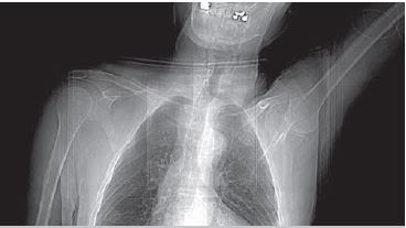
Shoulder CT Scan
Non contrast CT imaging is often requested of the evaluation of bony trauma in the shoulder. Thin slices are acquired in the axial plane beginning at the acromioclavicular joint and extending a few centimeters below the most inferior fracture line. This can be determined by carefully examination of the last few slices or asking a radiologist for assistance.
CT arthrography of the Shoulder
CT arthrography of the shoulder is useful for evaluation of the joint capsule and intracapsular structures and for finding loose bodies within the joint. CT Arthrography can be performed either with a single or double contrast technique. A 0.5 to 3 mL of iodinated contrast material and approximately 10mL of room air. Thin axial slices begin at just above the acromioclavicular joint and end just below the glenoid fossa.For either indication the patient is positioned supine on the CT table. The arm to examined is downward alongside the body, the opposite arm is extended over the patient’s head to reduce the xray beam absoption as much as possible.
CT arthrography of the shoulder is useful for evaluation of the joint capsule and intracapsular structures and for finding loose bodies within the joint.
For a shoulder CT the patient is positioned supine on the CT table. As seen on the image, the patients arm to be examined is downward, and the opposite arm is extended over the patient’s head to reduce the xray beam absorption as much as possible.


No comments:
Post a Comment