Radiologic Technologist Assessment and Monitoring of Vital Signs
Vital signs are the early indicators of a physiologic changes in a patient. Vital signs are body temperature, pulse, respiration and blood pressure. Radiologic technologist should begin to assess the patient when they first introduce themselves. It is important to notice the patient’s breathing, skin coloration, and overall health before the patient ever lies on the CT scan table. This will help the radiologic technologist notice signs should adverse effects occur during the scan process. Throughout the CT examination the patient should be monitored visually and spoken to frequently using the scanner’s intercom system. This reassures the patient and allows the technologist to intervene quickly when problems arise.Use of special monitoring devices
Special monitoring devices are not generally required for routine CT examinations performed on stable patients. However, patient from inpatient department may arrive in the CT department already connected to equipment such as monitors or a respirator. While you are in the CT department, unstable patient and the equipment must be watch carefully when they arrived. Because the CT scan technologist must focus on the task necessary to perform a high quality examination, a nurse or other health professional with appropriate training, should accompany these patients to provide the necessary monitoring and intervene should the need arise.Contrast Agents Adverse Reaction
It is obvious that many patients cared for in the Ct scan department are quite ill. In addition to the medical symptoms that necessary in the examination, patient may also have an adverse reaction to the IV contrast media used for the examination. Adverse effects are for the most, random and totally unpredictable. Therefore, radiologic technologist must be alert for physiologic changes in their patients. The best early indicators of a problem are changes in body temperature, pulse, respiration and blood pressure. Collectively these are called the vital signs or cardinal signs. The normal range of vital signs may vary somewhat with age, sex, weigh, exercise tolerance and patient condition. Other important indicators include pain, pulse oximetry values this is indicates blood oxygenation, and pupil size, equality and reactivity.Body Temperature and Monitoring Devices
Body temperature is most often taken by placing the thermometer in the mouth, the ear (using a tympanic infrared thermometer), the axilla or in the rectum. In CT scan department, the thermometer is most often an electronic, battery operated device with disposable protective sheaths. Other options are tympanic thermometer – also have with disposable sheaths.- Disposable single use chemical strip thermometers (such as 3M tempa-Dot),
- Mercury-free, glass thermometer – a blue tip typically denotes an oral thermometer where as red tip denotes a rectal thermometer
- Oral, rectal and tympanic thermometer – the temperature measurements are higher than axillary measurements because the measuring device is in contact with the mucous membrane.
Pulse
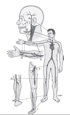
Each time the heart contracts if forces blood into an already full aorta. The elasticity of the arterial walls allows them to expand to accept the increase in pressure. Pulse is defined as the alternate expansion and recoil of an artery. By counting each expansion of the arterial wall in a given time frame the pulse rate can be determined. In general, the pulse can be felt wherever a superficial artery can be held against firm tissue, such as a bone.Some of the specific locations where the pulse is most easily felt are as follows:
a. Temporal Pulse or Superficial Temporal Artery – it is located just anterior to the ear.
b. Facial Pulse or Facial Artery – the lower margin of the mandible, about one third anterior to the angle.
c. Carotid Pulse or Carotid Artery – it is along the anterior aspect of the neck, to the right or left of midline.
d. Radial Pulse or Radial Artery – it is located at the thumb side of the wrist.
e. Brachial Pulse or Brachial Artery – it is located on the medial side of the elbow cavity, located between the biceps and triceps muscle, frequently used in place of the carotid pulse in infants.
f. Femoral Pulse or Femoral Artery – it can be found in the groin
g. Popliteal Pulse or Popliteal Artery – it is in behind the knee.
h. Pedal Pulse or Dorsalis Pedis Artery – it can be found on the top of the foot.
 |
| Locations where pulse can be taken. |
Blood Pressure Affects Pulse Palpability
The patient’s blood pressure will impact the ease of palpability of a pulse.
- If his systolic blood pressure is less than 90 mm Hg, the radial pulse will not be palpable.
- Less than 80mm Hg, the brachial pulse will not be palpable.
- Less than 60mm Hg, the carotid pulse will not be palpable. This is because systolic blood pressure rarely drops that low, the lack of a carotid pulse usually indicates cardiac arrest.
Pulse Average / Normal Ranges
The average adult pulse rate ranges from 60 to 100 beats per minute. However, in athletic adults, a normal pulse rate can range between 45 to 60 beat per minute. The average pulse rate for a child ranges from 95 to 110 beats per minute; infants from 100 to 160 beats per minute. When the pulse is being counted, the rate, rhythm, and volume should be noted. An irregular pulse is one that has a period or normal rhythm broken by periods of the pulse is often describe as full and bounding if it seem regular and with good force, or if is difficult to palpate and irregular it is often described as weak or thread.Respirations
The respiration rate is the number of breaths a person take per minute. It is usually measured when the patient is at rest and simply involves counting the number of breaths for 1 minute by counting how many times the chest arise. Normal respiratory rates vary according to age. Commonly accepted normal ranges are as follows:
- Adults – 14 to 20 Respiration / min
- Adolescent Youth – 18 to 22 respiration / min
- Children – 22 to 28 Respiration / min
- Infants – 30 or greater Respiration / min.
The ratio of respiration to pulse is fairly constant at approximately 1 breath to 4 heart beats.


No comments:
Post a Comment