Wrist CT Imaging
CT examination of the wrist are indicates for complex fractures involving the distal radius and ulna, scaphoid fracture, or other carpal fracture that are unclear on conventional xray images. CT may also be of value in the detection of subtle fractures.Patient Positioning
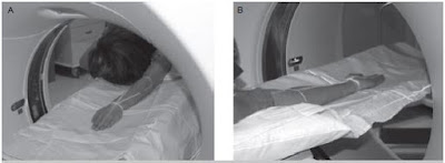
It is often difficult to comfortably and stably position the patient for a CT examination of the wrist. Different approaches are used. Sometimes the patient is positioned with his arm over his head. Another approach is to have the patient sit or stand on the far side of the scanner and extend his arm into the scanner. A 3rd, and less favorable, approach is to scan the patient’s wrist as it rests on his abdomen.Stable positioning is the key to obtain a good image quality by avoiding motion artifacts. However, patient positioning of the wrist is at best awkward and particularly in patients with other trauma related injuries, can pose a significant obstacle to the study. Many different approaches are used:
The most common probable technique is to extend the patient’s arm over the head. This can be done with the patient lying supine or prone.
 |
| Patient lie on the scanning table, either prone or supine |
Another approach is to have the patient sit or stand at far end of the CT scanner with his arm resting on the CT table.
 |
| Patient sit or stand at the far end of the CT scanner |
Direct Coronal Wrist CT Imaging
If direct coronal imaging is desired, the patient’s elbow is bent so that his arm is positioned parallel to the gantry. The patient is told to maintain the position while the table moves a short distance and is instructed to move his body along with the table. The patient is asked to look away from the gantry to minimize corneal radiation exposure. If the patient’s hand cannot be positioned using one of these approaches, a third method is to position the patient supine on the table with his arm resting on the belly. Scans are obtained through both the wrist and the section of the abdomen on which it rests. In this situation tube current must be increased to at least 300mAs so that image noise is sufficiently suppressed. Because of the radiation exposure to the patient’s abdomen, this method is used only when other approaches have failed.
Wrist CT Anatomy
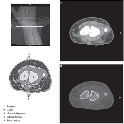
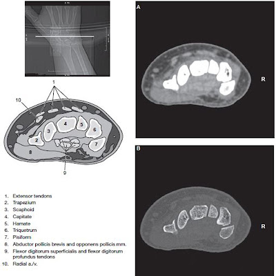
Proper positioning and appropriate annotation of the elbow, wrist, forearm and hand can be challenging. Confusion as to whether an extremity is the right or left may occur when the patient’s arm is positioned over the patient’s head. Therefore, it is common practice for the radiologic technologist to include a radiopaque marker to aid in identification of the scan orientation. In the image below notice the BB marker is placed on the ulnar side of the right wrist.
 |
| Wrist CT Scan |


No comments:
Post a Comment