Skull Radiography : Positioning Projections and Methods
Cranium
Projections / Views
- Lateral Projection : Rotated Right or Left
- Dorsal Decubitus Projection : Right or Left Laterals
- PA (Posteroanterior) View
- AP (Anteroposterior) View
Radiographic Methods of the Skull
- Caldwell Method - PA Axial Projection
- Towne Method - AP Axial Projection
- Haas Method - PA Axial Projection
X-ray Examination of the Cranial Base of the Skull
Radiographic Methods
- Submentovertical (SMV) – Schuller Method
- Verticosubmental (VSM) – Schuller Method
 |
| Anterior View |
Skull Anatomy and Pathology
The skull is located on the superior part of the vertebral column. There are 22 separate bones these are divided into 8 cranial bones and 14 facial bones. The cranial bones are further divided into the calvaria and floor.The cranial bones protects and acts as the housing of the brain. Facial bones provides structure, shape and support for the face and forms the protective housing for the upper ends of the respiratory and digestive tracts plus with several protection for the structures and organs of our sight.
The 22 primary bones of the skull should be located and recognized in the different views before they are situated in greater detail. The inner layer of the cranial vault is separate by spongy tissue (diploe) and composed of 2 plates of compact tissue, outer plate or table is thicker than the inner table. Inner layer of cranium (spongy tissue) varies considerably.
 |
| Lateral View |
Cranial Bones Compositions
Calvaria1 Frontal
1 Occipital
1 Right parietal
1 Left parietal
Floor of the Skull
1 Ethmoid1 Sphenoid
1 Right temporal
1 Left temporal
 |
| Superior View |
Facial Bones Composition
2 Nasal
2 Lactimal
2 Maxillary
2 Zygomatic
2 palatine
2 Inferior nasal conchae
1 Vomer
1 Mandible
Sutures of the skull
The sutures are fibrous joints that connects the cranium and the face.Coronal – it is found between the frontal and parietal bones.
Sagital – it is located on the top of the head between the two parietal bones and just behind the coronal suture line
Squamosal – It is between the temporal bones and the parietal bones.
Lambdoidal – Between the occipital bone and the parietal bone.
Junction of the coronal and sagittal sutures.
Lambda – junction of the lambdoidal and sagittal sutures.
Bregma – it joints the coronal sutures and sagittal sutures.
Pterion - Junction of the Parietal Bones, Squamosal and Greater Wing of the sphenoid, and lies in the middle of meningeal artery.
Asterion - Junction of the occipital bone, parietal bone and mastoid portion of the temporal bone.
Common Radiographic Pathology of the Skull
 |
| Anterior View Landmark |
- Fracture – Disruption in the continuity of the bone
- Basal – A fracture located at the base of the skull
- Blowout – Fracture of the floor of the orbit
- Contre-coup – Fracture to one side of a structure caused by trauma to the other side.
- Depressed – Fracture causing a portion of the skull to be depressed into the cranial cavity.
- Leforte Fracture – Bilateral horizontal fractures of the maxillae
- Linear Fracture – Irregular or jagged fracture of the skull
- Tripod Fracture – Fracture of the zygomatic arch and orbital floor or rim and dislocation of the frontozygomatic suture.
- Mastoiditis – Inflammation of the mastoid antum and air cells.
- Metastases – Transfer of a cancerous lesion from area to another.
- Osteomyelitis – Inflamation of bone in the skull due to a pyogenic infection.
- Osteopetrosis – Increased density of atypically soft bone.
- Osteoporosis – Loss of bone density.
- Paget’s Disease – Thick, soft bone marked by bowing and fractures.
- Polyp – Growth or mass protruding from a mucous membrane
- Sinusitis – Inflamation of one or more of the paranasal sinuses.
- TMJ Syndrome – Dysfunction of the temporomandibular joint.
- Tumor – New tissue growth where cell proliferation is uncontrolled.
- Acoustic Neuroma – Benign tumor arising from Schwann cells of the eight cranial nerve.
- Multiple Myeloma – Malignant neoplasm of plasma cells involving the bone marrow and causing destruction of the bone.
- Osteoma – Tumor composed of bone tissue.
- Pituitary Adenoma – Tumor arising from the pituitary gland, usually in the anterior lobe.
 |
| Lateral View Head Landmark |
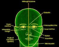
- Midsagittal Plane
- Interpupillary Line
- Acanthion
- Outer Canthus
Infraorbital Margins
- External Acoustic Meatus (EAM)
- Orbitomeatal Line (OML)
- Infraorbitalmeatal Line (IOML)
- Acanthiomeatal Line (AML)
- Mentomeatal Line (MML)
The skull of adult has a normal of 7° angle difference existence between the OML and IOML, and a difference of 8° angle between the OML and the glabellomeatal line. The difference in degree of angle between the cranial lines used in positioning must be known. Often the relationship of the patient, Image receptor, and central ray is the similar, but its angulation has described differently, depending on the lines of the cranial references.
Cranium


